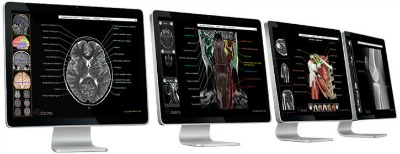
![]() VisualDX is a visual diagnostic decision support system that includes medical images and illustrations. VisualDx is designed to match the way a physician thinks about signs, symptoms, and diagnoses. The content is organized by common and diagnostic problem areas as well as by age and body location. It visually presents disease variations by skin type, age, and passage of time. Clinicians can initiate a unique differential diagnosis based on the visual symptoms of a patient. A customized pictorial differential diagnosis tailored to a patient’s findings would be formulated dynamically. VisualDX also compares most relevant diseases, or drills down to research with handbook-length clinical information and variation in visual presentation.
VisualDX is a visual diagnostic decision support system that includes medical images and illustrations. VisualDx is designed to match the way a physician thinks about signs, symptoms, and diagnoses. The content is organized by common and diagnostic problem areas as well as by age and body location. It visually presents disease variations by skin type, age, and passage of time. Clinicians can initiate a unique differential diagnosis based on the visual symptoms of a patient. A customized pictorial differential diagnosis tailored to a patient’s findings would be formulated dynamically. VisualDX also compares most relevant diseases, or drills down to research with handbook-length clinical information and variation in visual presentation.
To access this resource, click on the link above, or look it up using the MSKsearch search box on the Library website.
From Your Mobile Device (for iOS and Android only):
MSK clinical staff can access VisualDx on their mobile device due to an institutional subscription to this resource. Follow the four easy steps below to download the app:
- Log in to VisualDx on a computer at the hospital, through VPN, or from the Library Website
- Find the VDx Mobile icon (upper right corner) and click the Get VisualDx Mobile link
- Using your institutional e-mail address, complete the form to request a user name and password
- Follow the instructions in the activation e-mail to download and begin using VisualDx Mobile
Please note that you will need to request a user name and password in order to access the app. Should you need assistance, please contact the reference team via Ask-A-Librarian or 212-639-7439.
Access to this resource has been made possible by the Department of Medicine, Dermatology Service. Their financial support is greatly appreciated.